




Glaucoma in dogs and cats
Glaucoma is an eye disease affecting the optic disc and certain retinal cells, the ganglion cells. These structures transmit information gathered by the photoreceptors to the optic nerve, so that dysfunction leads to reduced visual acuity or even blindness. This disease, known in humans, also affects dogs and cats.
How to recognize glaucoma
Unlike in humans, glaucoma in carnivores usually develops acutely. It manifests itself as ocular redness, pupil dilation (mydriasis) and sometimes a reduction in corneal transparency (corneal edema). From a functional point of view, affected subjects experience pain that manifests itself (but not always) as eyelid spasm (blepharospasm) and a marked drop in visual acuity, or even total loss of vision on the affected side. The veterinarian confirms the presence of glaucoma by observing these characteristic clinical and functional signs, and by identifying an increase in intraocular pressure (known as ocular hypertonia), which he measures with a tonometer.

Why does my dog have glaucoma?
The entire anterior part of the eye (anterior segment) is filled with a liquid called aqueous humor. This fluid is involved in ocular tone. It is constantly produced by the ciliary processes and at the same time evacuated by drainage structures contained in the iridocorneal angle. The eye can thus be compared to a balloon, constantly supplied by several taps (ciliary processes) and drained in parallel by a siphon (iridocorneal angle) located around its entire periphery. The ocular hypertonia observed in glaucoma is the consequence of a defect in aqueous humor drainage. If we refer to the balloon model, liquid is produced by the valves, but the siphon no longer performs its draining function properly, resulting in a rise in balloon pressure.
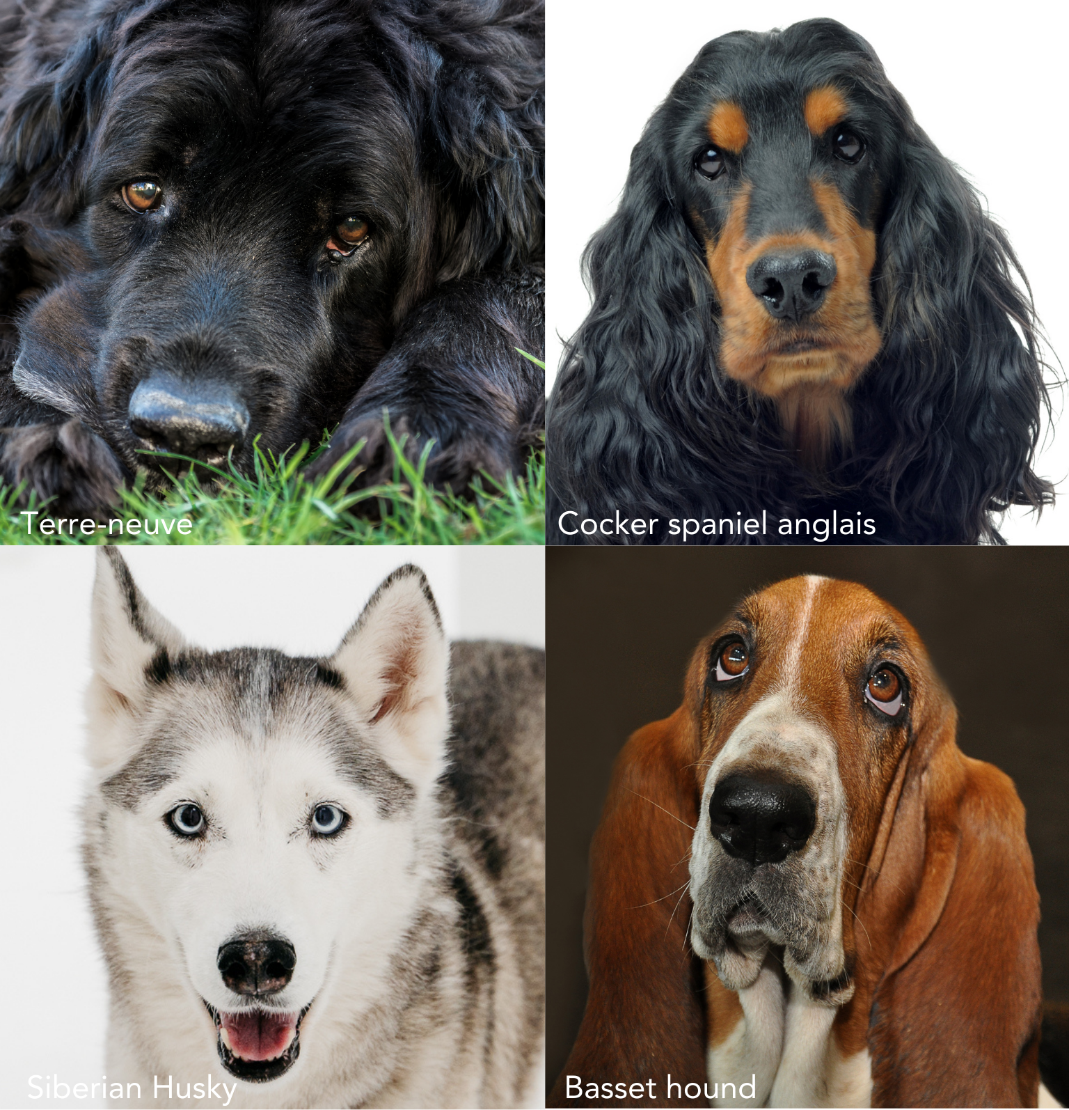
There are two types of glaucoma. Primary glaucoma due to a structural anomaly of the iridocorneal angle. This anomaly is most often bilateral. Certain breeds, such as the Cocker Spaniel, Bouvier des Flandres, Basset Hound, Siberian Husky, Newfoundland, Shar Pei and Labrador, are predisposed. This list is not exhaustive. Secondary glaucoma due to acquired obstruction of the iridocorneal angle. In this case, glaucoma is the consequence of another ocular disease (uveitis, tumor, cataract, lens dislocation, etc.) that disrupts the evacuation of aqueous humor.
In short, there are glaucomas due to structural causes linked to the patient's anatomy; they appear during the dog's life as the aqueous humor drainage pathways no longer fulfill their role, and acquired glaucomas, secondary to another ocular disease that occurs during the subject's life.
What are the consequences of glaucoma?
The rise in intraocular pressure that accompanies glaucoma causes damage to ganglion cells, neuroretinal fibers and the optic disc. These highly sensitive structures are responsible for transmitting visual information. When these structures are damaged, vision is affected, and the subject can very quickly lose his or her sight. The higher the level of ocular hypertonia, the more serious the damage. If it persists, the damage is irreversible.
What's more, increased pressure in the eyeball can cause acute pain.
In this respect, acute glaucoma is an emergency that needs to be treated rapidly in order to preserve visual function.

What treatment options are available?
The mainstay of treatment for carnivore glaucoma is to counteract the increase in intraocular pressure. There are two categories of treatment:
- Medical treatment with local medications (anti-glaucoma eye drops) and systemic medications. These drugs have a variable effect on intraocular pressure, and do not always reduce it sufficiently to avoid blindness and pain. While eye drops are generally well tolerated, despite a few local reactions, systemic medications have side effects that make them unsuitable for long-term use.
- Surgical treatment consists of either reducing aqueous humor production by destroying part of the ciliary processes - known as cyclo-weakening - or improving aqueous humor evacuation by creating an evacuation pathway - known as fistulizing surgery.
Surgical techniques are the alternative of choice when medical treatment is unable to control ocular hypertonia on a lasting basis. Unfortunately, however, they do not provide an infallible solution to glaucoma in all cases, and can sometimes be accompanied by recurrence or failure. To increase the success rate, it is crucial to treat glaucoma patients as early as possible, and in particular before the onset of blindness, which in most cases is likely to be permanent. In this respect, acute glaucoma is an ocular emergency.
What does the assessment of a glaucoma patient include?
When treating glaucoma patients, it is important to determine the origin of the ocular hypertonia. This step is crucial to choosing the right treatment. Is it a cataract causing the glaucoma, in which case surgery is required? Is it a primary dislocation of the lens, also requiring surgery? Is it primary glaucoma, in which case there is a significant risk of glaucoma in the other eye? Is there a risk of lens dislocation in the other eye? These are just some of the questions that need to be answered in the assessment of a glaucoma patient. This assessment involves an exhaustive clinical examination of both eyes, carried out by an experienced veterinarian. It also includes an examination of the aqueous humor drainage structures (gonioscopy, ultra-biomicroscopy or UBM) and, when useful, of the optic disc (optical coherence tomography or OCT).
