




MRI IN VETERINARY MEDICINE
MRI
Magnetic Resonance Imaging (MRI) is an advanced medical imaging examination, essential for exploring soft tissues, the nervous system and internal structures in animals. This cutting-edge technology provides detailed and precise images, facilitating the diagnosis of many complex pathologies.
Technical advances have made MRI an indispensable tool in neurology, orthopedics, oncology and veterinary internal medicine. It offers a fine visualization of the brain, spinal cord, joints and internal organs, enabling precise identification of structural and functional abnormalities.
The Agoravet veterinary clinic is one of the few clinics in France to have a 1.5 Tesla high-field MRI, offering optimum imaging quality for accurate diagnoses. Thanks to our expertise in advanced imaging and close collaboration with medical and surgical departments, we are able to provide comprehensive, tailored care for animals requiring in-depth examinations.
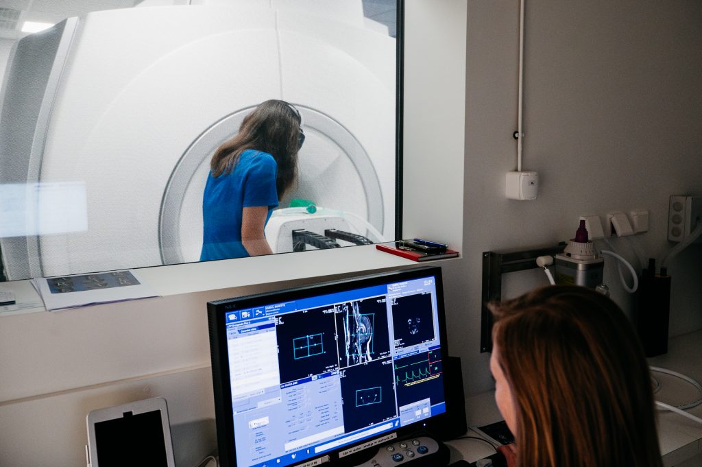
How it works
What examinations are possible with veterinary MRI?
Brain and spinal cord MRI
MRI is the gold standard in veterinary neurology. It provides detailed images of the brain and spinal cord, essential for diagnosing complex pathologies such as :
- Brain and spinal cord tumors
- Herniated discs and other spinal injuries
- Encephalitis and meningitis
- Strokes and other ischemic strokes
- Hydrocephalus and other congenital malformations
- Fibrocartilage embolism
This examination is particularly recommended for animals with neurological disorders, such as convulsions, sudden paralysis or loss of balance.
Joint and musculoskeletal MRI
MRI enables precise exploration of ligaments, cartilage, muscles and joints. It is used for :
- Ligament ruptures( anteriorcruciate , tendonitis, etc.)
- Muscular and tendon pathologies
- Complex spinal fractures and associated spinal cord injuries
Thanks to MRI, it is possible to make a precise diagnosis and adapt treatment, whether surgical or medical.
MRI of soft tissues and internal masses
MRI is a key tool for assessing tumors, cysts and deep inflammation. Unlike other imaging techniques, it enables detailed analysis of the structure of masses and evaluation of their extension. It is particularly indicated for :
- Exploring brain and spinal tumors
- Assessment of suspicious abdominal or thoracic masses
- Analysis of muscular or peripheral nerve inflammation
MRI thus enables us to refine the diagnosis and better plan any surgical intervention.
MRI in ophthalmology and ENT
Certain ophthalmological or otolaryngological pathologies require in-depth examination. MRI is used to diagnose :
- Retrobulbar abscesses and other orbital disorders
- Tumors of the sinuses, nasal cavities and inner ears
- Deep upper respiratory tract infections
These conditions can be difficult to detect using conventional tests. MRI provides a detailed analysis of the structures affected, for optimized management.
Care and maintenance
How can I have an MRI performed at the Clinique Agoravet in Strasbourg?
Your veterinarian may recommend an MRI if conventional imaging tests (X-rays, ultrasounds, scans) do not provide a precise diagnosis. In this case, you should make the request directly on the AgoraVet website.
MRI examinations are carried out by appointment only, under general anesthesia to guarantee the immobility required for the examination. The Agoravet team provides constant anesthesia monitoring and rigorous post-examination follow-up to ensure your pet's safety. It is essential that your pet does not eat or drink anything.
Images obtained during MRI examinations are systematically sent to veterinary imaging specialists for detailed interpretation. This expert analysis ensures a precise diagnosis and optimizes the management of each animal. This process may require additional time before the final report is sent.
The collaborative team
Dr. Olivier Guillaume
Dr. Roxane Lebel
Melissa
Davina
Dr. Xavier Ferreira
Dr. Audrey Hranitzky
Dr. Tess Gauthier
Dr. Séverine Hafner
Service news
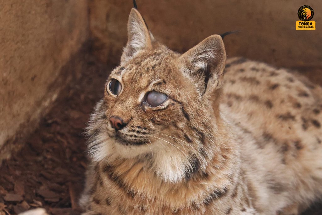
A lynx taken care of at Agoravet
Some stories are truly memorable, and this is one of them. A young female lynx has been presented to our […]
Read more
Case review: Bilio-pancreatic MRI
Anamnesis: Chronic recurrent pancreatitis. MRI sequences : The study consists of several series of MRCP-weighted 3D lava images of the […]
Read more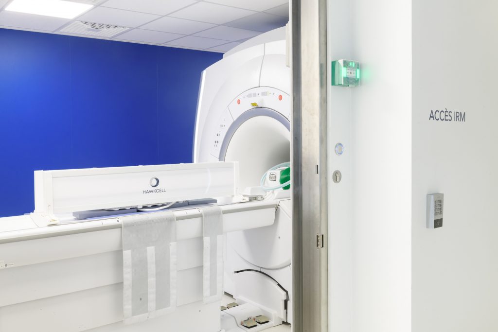
Case Review: Otitis
Anamnesis: Dog presented with balance disorder, head tilted to the right, ptyalism, ear sensitivity and horizontal nystagmus at rest. MRI […]
Read more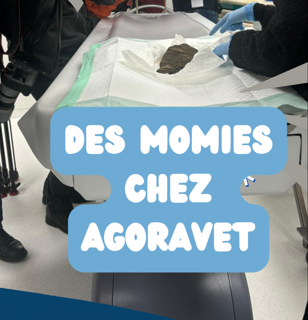
It happened at the clinic! 🎥📸
An unusual operation took place at the clinic. Researchers from the University of Strasbourg used our equipment to scan five […]
Read more